Intraoral Cameras: Catching Oral Health Issues Earlier
Most oral health problems don’t announce themselves. A cavity forming between two teeth, a patch of irritated tissue on the inside of a cheek, the early signs of gum recession, these are easy to miss in a mirror, and to overlook until they have already progressed. Intraoral cameras were built to close that gap. By magnifying and documenting what’s happening inside the mouth, they give patients and providers a shared, visual record instead of a rushed verbal description.
Below, we look at what the research and clinical experience actually show about intraoral camera use, where it helps most, what the latest studies say about patient comfort, and why at-home imaging is becoming part of that picture.
What an Intraoral Camera Actually Shows you
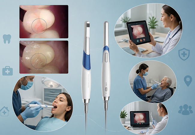
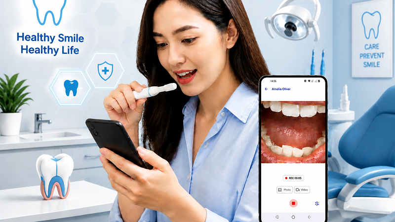
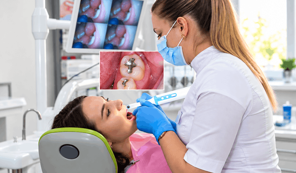
An intraoral camera is a small, pen-sized camera, often paired with LED lighting and magnification, that captures close-up images or video of teeth, gums, and soft tissue. In a dental office, a hygienist or dentist typically guides the camera during an exam. At-home devices like MouthCAM extend that same idea to the patient: a wireless or wired camera that connects to a phone or computer, so imaging can happen between visits rather than only during them.
The output isn’t just a picture. It becomes a dated, comparable record — something a provider can set next to an image from six months earlier to see whether a spot has changed, grown, or resolved on its own.
Why Early Detection Depends on Documentation, Not Just Inspection
Oral cancer screening is one of the clearest examples of why this matters. More than 54,000 Americans are diagnosed with oral cancer each year, tobacco use, alcohol use, and HPV being the most common risk factors. Dental industry guidance on screening points to a specific, practical use for intraoral imaging: establishing a baseline record of soft tissue, then re-checking it at future visits to catch changes early.
That documentation does two things. First, it makes it possible to notice a lesion that’s grown or changed color since the last visit, something that’s hard to judge from memory alone. Second, when a provider does need to refer a patient to a specialist, a clear image gives the referral more clinical weight than a written note describing what was seen. Additionally, it provides the patient with something tangible to look at, which tends to strengthen the argument for following through on that referral more than a verbal warning alone..
The same logic applies below the gumline. Cavities and early gum disease are also easier to miss on a quick visual exam than on a magnified image, which is part of why intraoral cameras have become an essential part of many exams even outside of cancer screening specifically.
Seeing the Problem Changes the Conversation
Dental offices that use intraoral cameras regularly report a second, less clinical benefit: patients understand their own treatment plans better. One practice described a patient’s experience this way , the camera “made my visit informative and engaging” rather than something to sit through passively.
That tracks with a broader pattern in patient communication. It’s one thing to be told there’s early decay between two molars. It’s another to see the dark spot on a screen, in your own mouth, magnified. Visual evidence tends to reduce the back-and-forth of “are you sure?” and replace it with an informed decision, whether that means agreeing to a filling now or understanding exactly what to watch for later.
What the Latest Research Says About Comfort
Newer imaging isn’t automatically more comfortable imaging, and it’s worth being honest about that. A 2026 observational study conducted with 192 participants compared intraoral camera exams to smartphone-based photography for dental imaging. It found that comfort decreased for about 1 in 7 participants (15%) during a traditional intraoral camera exam, a statistically significant drop. Smartphone imaging didn’t show that same overall decline though the study also found that younger participants (ages 6 to 19) were actually more likely to find smartphone imaging uncomfortable than adults were, at 38% versus 10%.
The researchers’ takeaway wasn’t that one method is universally better. It was that comfort varies by device, by age group, and by what’s being imaged and that patient-friendly alternatives are worth continued study, particularly for reaching people who don’t have easy access to a dental office.
That study looked specifically at a clinician performing the imaging during an in-office visit. It didn’t test what happens when a patient captures their own images, at home, at their own pace. That’s a meaningfully different experience: no one else’s hands are in your mouth, there’s no clinical setting to feel rushed in, and you control when and how the camera is used. It’s a reasonable expectation, though not something the study itself measured, that self-directed imaging removes some of the specific discomfort the study identified.
Who Gets the Most Value From At-Home Imaging
Between-visit imaging tends to matter most for a few groups in particular:
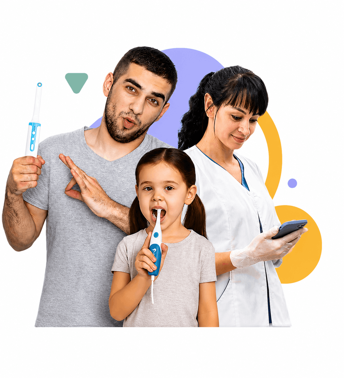
Parents monitoring a child’s teeth as they come in or shift can flag a concern, days or weeks before the next scheduled cleaning, rather than waiting and hoping it doesn’t get worse.
Patients managing dental anxiety often find it easier to look at their own mouth on their own terms than to sit through an in-office camera exam, and a home image still gives their dentist something concrete to review remotely.
People in rural areas or without easy access to regular dental care can use imaging as part of a teledentistry visit, sending a provider a clear picture instead of describing symptoms over the phone.
Adults tracking a specific issue, a receding gumline, a chipped filling, a spot that a dentist asked them to “keep an eye on” get an actual record to compare over time, instead of relying on memory.
The Bottom Line
Intraoral cameras don’t replace a dentist’s judgment, and no camera in-office or at-home substitutes for a professional exam when something looks wrong. What they add is documentation: a comparable, shareable record that supports earlier detection, clearer referrals, and better-informed patients. Combine that with the growing evidence that comfort and accessibility matter just as much as image quality, and the case for imaging that patients can do themselves, between visits, becomes hard to ignore.